NEW YORK (Reuters Health) – In patients with suspected pulmonary embolism (PE), computed tomographic angiography (CTA) is more than twice as likely to show an unexpected nodule or adenopathy as it is to show a PE – and physicians need to be prepared to deal with the consequences, warns a paper in the November 23 issue of Archives of Internal Medicine.
The burdens of such incidental findings “are not inconsequential,” the authors add. Follow-up tests are likely to involve significant amounts of ionizing radiation, patients are subjected to the stress of being told they may have a malignancy, and clinicians must spend a considerable amount of time explaining the test result and the recommended follow-up.
All of this must be added to the actual cost of follow-up studies and related complications, the investigators note.
“As pulmonary consultants, we have been struck by the increasing number of referrals to evaluate pulmonary nodules and adenopathy first detected on CTAs,” principal investigator Dr. Shannon S. Carson, from the University of North Carolina at Chapel Hill, told Reuters Health. “The indications for many of those CTAs were not always clear, and they were ordered almost as a screen for respiratory symptoms. This approach to diagnostic testing could of course lead to a high rate of incidental findings.”
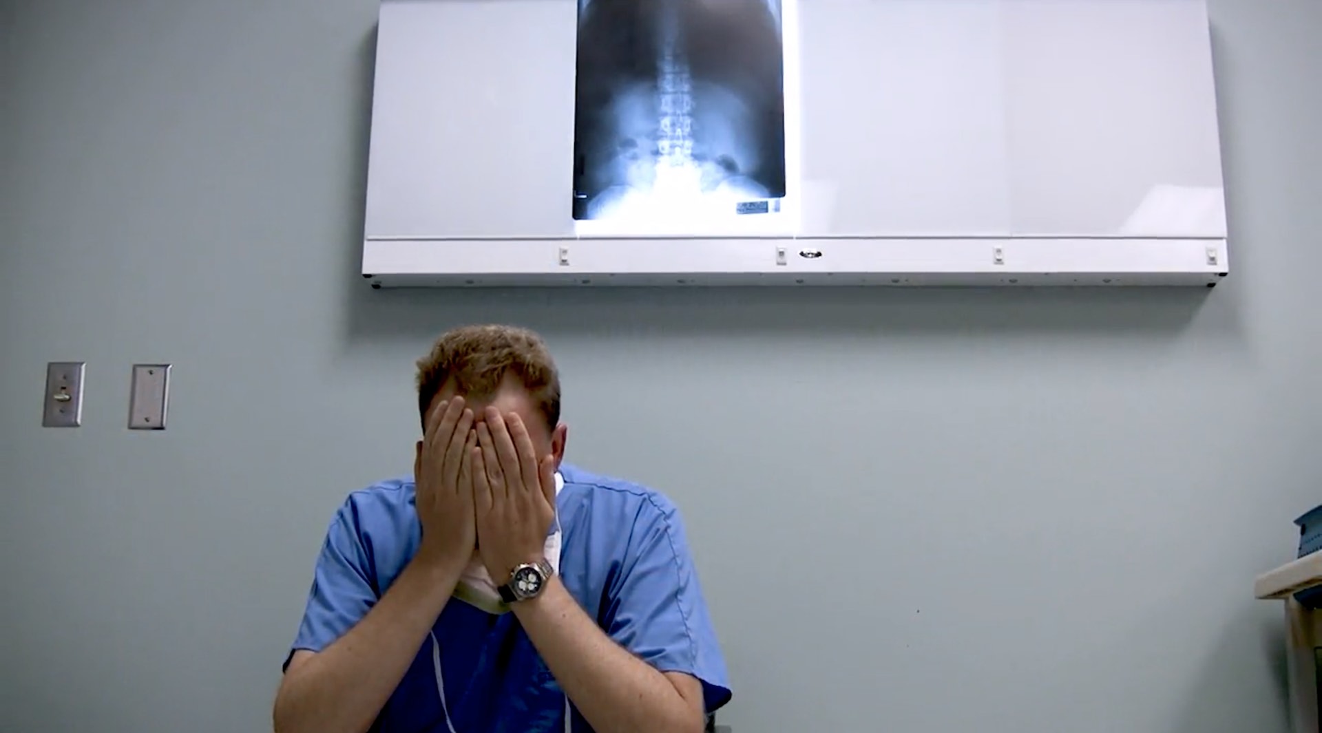
In their single-center study, Dr. Carson and her associates reviewed 589 CTAs that had been ordered in the emergency department. In 55 cases (9%), CTA confirmed PE. In 141 patients – nearly a quarter of those tested — CTA showed an incidental finding that required diagnostic work-up.
Seventy-three patients (13%) had a new pulmonary nodule (a mass of less than 3 cm) in or arising from the pulmonary parenchyma. Fifty-one patients (9%) had a new incidental adenopathy.
“Using current clinical guidelines, follow-up CT or another procedure would be recommended for 96% of patients with new incidental pulmonary nodules” in this study, the authors write. They add that formal guidelines for follow-up of incidental pulmonary adenopathy don’t exist “and may need to be established.”
“To avoid unnecessary, burdensome, and low-yield follow-up studies, systematic approaches to determining clinical risk and therefore higher yield indications for CTA are recommended during assessments of acute pulmonary symptoms in the emergency department,” the investigators conclude.
Dr. Carson recommends the following: “Except in patients presenting with severe shock needing immediate decisions regarding thrombolytic therapy, a good-quality chest radiograph should be obtained in all cases before moving on to CT angiogram. If major risk factors are absent, and alternative diagnoses are more likely based upon history, physical exam, chest radiography, ECG, and blood tests such as a D-dimer level, then a CT angiogram should not be ordered.”
“This will improve the positive predictive value of CT angiogram and reduce needless follow-up of incidental findings,” she added.
Reference:
Arch Intern Med 2009;169:1961-1965.