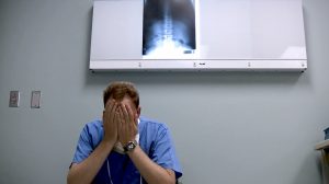
NEW YORK (Reuters Health) – Multidetector CT (MDCT) identifies skeletal chest injuries after cardiopulmonary resuscitation (CPR) missed by conventional x-rays, researchers from Korea report in the June 12th online Resuscitation.
Rib and sternum fractures frequently complicate thoracic compression administered during CPR, but this is the first study to evaluate MDCT findings of such chest injuries as compared with the findings of radiography.
Dr. Hyuk Jun Yang and colleagues from Gachon University Gil Hospital, Incheon, Republic of Korea compared MDCT and radiography findings in 40 patients who underwent successful CPR within the preceding 24 hours.
MDCT identified 173 rib fractures in 26 patients and 12 sternal fractures. Radiography detected rib fractures in only 10 patients and failed to detect any sternal fractures.
Most of the cases had multiple, bilateral rib fractures, and most sternal fractures occurred in the middle and lower third of the sternum.
Six patients had rib fracture-related complications, including chest wall hematoma (4 patients), pneumothorax (1 patient), and subclavian vein injury (1 patient), and 6 patients showed secondary signs of sternal fracture (retrosternal or mediastinal hematoma).
“Chest radiography remains the primary screening study for assessing patients who have chest trauma,” the researchers conclude. “However, the use of MDCT has increased for these patients because it enables thinner sections with greater speed, and this allows higher quality axial images and multiplanar reconstructions.”
The authors stopped short of recommending MDCT in all patients who survive CPR, but emphasized that it “is useful for the evaluation of chest injuries secondary to CPR as compared with that of radiography and also for the evaluation of the fracture-related complications.”
Resuscitation.